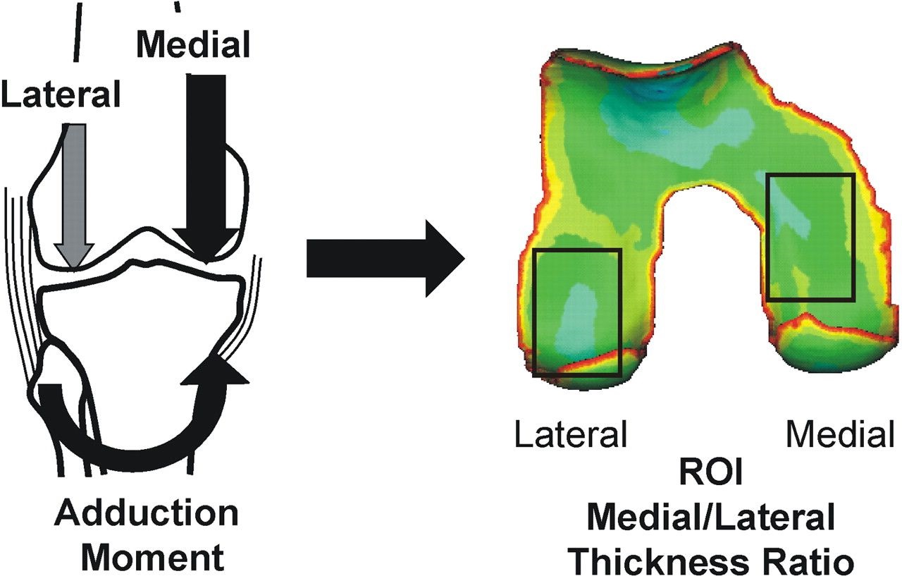
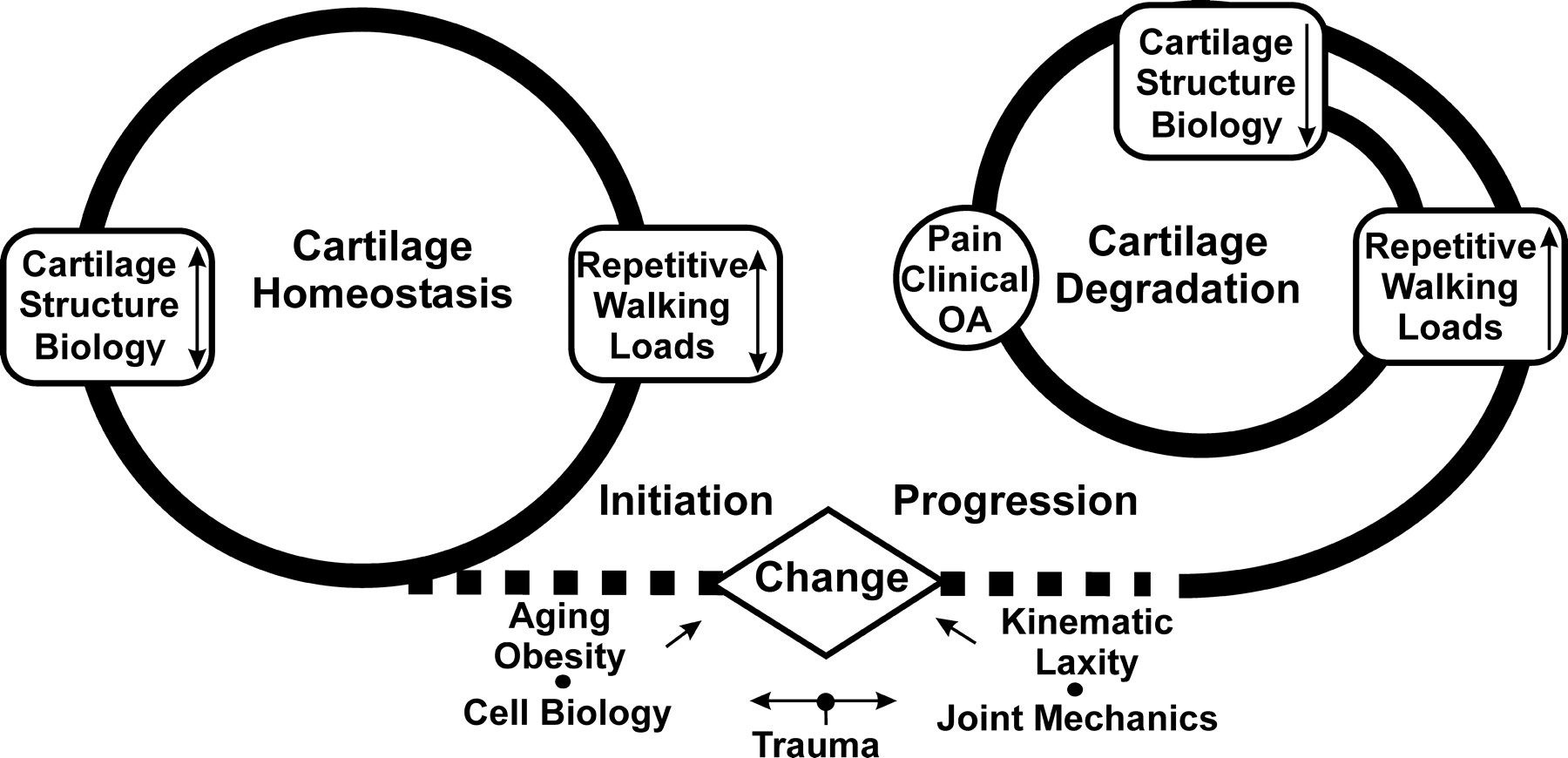
The question proposed by the current study is: Is there a combination of biomechanics, biochemistry, and MRI-detectible ACL biological composition which can cumulatively and accurately predict ACL injury risk? The long-term goals are three-fold. (1) determine a complete ACL injury risk profile by evaluating biomechanics, biochemistry, and ACL biological composition, (2) develop a useful screening protocol for at-risk athletes for the purpose of implementing a prevention/training regimen to reduce the overall risk of ACL injury, and (3) better characterize the success of ACL reconstructive surgery for restored biomechanics and joint health (i.e. osteoarthritis initiation) following injury. The figures below are from Andriacchi et al., 2009.


Diagram showing how knee adduction moment increases load on the medial compartment of the knee. On the left, arrows indicate higher medial loading and lower lateral loading. On the right, a color-mapped 3D representation of the distal femur shows cartilage thickness, with boxed regions marking medial and lateral areas used to calculate the medial-to-lateral cartilage thickness ratio.Flow diagram illustrating two parallel cycles: one representing cartilage homeostasis and the other cartilage degradation. The homeostasis cycle includes cartilage structure biology and repetitive walking loads. A central ‘Change’ box connects initiation factors such as aging, obesity, and trauma to progression factors like joint mechanics and kinematic laxity. The degradation cycle includes worsening cartilage structure biology, increased repetitive walking loads, and leads to pain and clinical osteoarthritis.