|
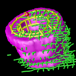
The data field generated by these images is rather sparse. In order to estimate the displacement field, a bounding box is placed around the image data, and then broken down into a mesh. A displacement vectors is then estimated for each point in the grid based on changes in the grid over time. The first step in the reconstruction is segmenting the myocardium based on the tag line data. In this step, the false tag points from the tag tracking stage are eliminated base on the spatial position of all the tag points in the dataset. Our algorithm is based on level sets. The resulting segmentation is shown in magenta. |
Because it cannot be known exactly where a specific tag line will displace from one frame to the next (the displacement in only a single direction of the three vector directions used can be known), the problem requires addtional assumptions about the nature of the myocardium being modelled. In essence, these assumptions are that the muscle fibers of the myocardium are connected in both the fiber and cross fiber direction, which is supported anatomically. These assumptions are modelled by a 3-D spatial smoothness constraint which can be incorporated into the displacement equation and allow it to be solved.
The strain field is calculated by calculating a stain tensor for each point beyond time frame zero based on the displacement field. The final results can then be exported into a visualization program for diagnosis.